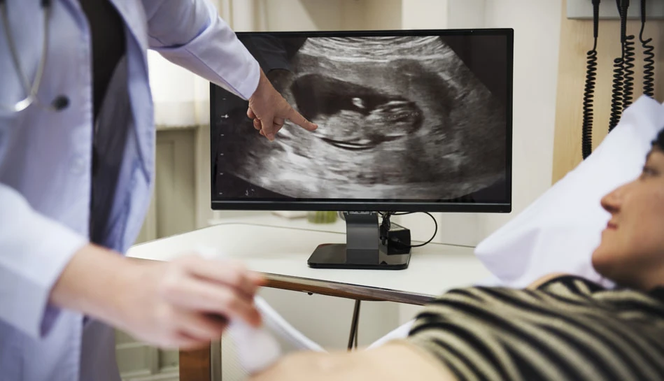
Fetal echocardiography is a specialized ultrasound examination used to evaluate the structure and function of a baby’s heart before birth. The fetal heart begins developing early in pregnancy, and detailed imaging helps doctors detect certain congenital heart conditions or abnormalities that may affect the baby’s health.
This scan provides a close assessment of the baby’s heart chambers, valves, and blood flow patterns. It helps identify potential cardiac concerns early so that parents and doctors can plan appropriate monitoring and care during pregnancy and after delivery.
At Fetal View Sonography Centre, fetal echocardiography is performed using advanced ultrasound technology and carefully interpreted by Dr. Pranita Mahashabde, a fetal medicine specialist with extensive training in prenatal diagnosis. With precise imaging and compassionate counselling, the focus is on providing clear information to expecting parents and ensuring the best possible care for both mother and baby.
The baby’s heart is one of the most important organs to evaluate during pregnancy. A dedicated fetal heart scan allows specialists to examine cardiac structures in detail and detect certain congenital heart conditions before birth.
Fetal echocardiography provides a comprehensive view of the baby’s heart. Doctors carefully examine the heart chambers, valves, blood vessels, and rhythm to ensure normal cardiac development.
Modern ultrasound techniques allow specialists to evaluate blood flow and heart function with high precision. Early identification of cardiac abnormalities allows timely monitoring and medical planning.

“Understanding the baby’s heart health during pregnancy helps doctors plan the safest care for both mother and baby. Early detection of cardiac conditions allows families to receive the right guidance and support throughout pregnancy.”
Fetal echocardiography helps doctors study several important aspects of the baby’s heart development.
The scan carefully examines the four chambers of the baby’s heart, valves, and major blood vessels to ensure that the heart is forming normally during pregnancy.
Using Doppler ultrasound, specialists assess blood flow through the fetal heart and major arteries to confirm healthy circulation between the heart and the rest of the body.
Fetal echocardiography evaluates the baby’s heartbeat and rhythm to detect possible irregular heart patterns or rhythm abnormalities.
This scan helps detect congenital heart defects early in pregnancy, allowing doctors to plan appropriate monitoring and care before and after birth.
The examination also helps doctors evaluate how blood circulates between the placenta and the baby, which is important for overall fetal well-being.
Fetal echocardiography may be recommended for pregnancies with higher risk factors, such as family history of heart disease, maternal diabetes, or abnormal findings on routine pregnancy scans.
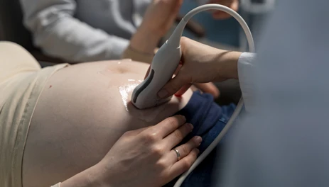
Fetal echocardiography is usually performed during the second trimester when the baby’s heart structures can be clearly visualized.
A detailed ultrasound examination is performed to evaluate the baby’s heart anatomy and function.
Specialists carefully study the chambers, valves, and blood flow patterns of the fetal heart.
Parents receive a clear explanation of the findings and guidance on any further monitoring or care if needed.
Fetal echocardiography not only helps evaluate the baby’s heart health but also provides reassurance for parents during pregnancy.
Advanced ultrasound technology is used to obtain detailed images of the fetal heart.
The scan is carefully analyzed by a fetal medicine specialist trained in prenatal cardiac evaluation.
Parents receive clear explanations and recommendations based on the scan results.

All scans are performed under the supervision of Dr. Pranita Mahashabde, a fetal medicine specialist with advanced expertise in prenatal diagnosis, ensuring accurate evaluation and compassionate care for every pregnancy.
Trustindex verifies that the original source of the review is Google. I had a wonderful experience during my anomaly scan with Dr. Pranita. My baby was being shy and not revealing the face, but she was incredibly patient and kind throughout the process. Instead of rushing through, she asked me to wait and took the time needed to get all the important details. Her calm and cooperative nature made me feel truly cared for. I'm very grateful for her professionalism and compassion. Highly recommend her to any expecting motherPosted onTrustindex verifies that the original source of the review is Google. Supportive & positive ambience with lower rates as compared to market. Must recommended.Posted onTrustindex verifies that the original source of the review is Google. Posted onTrustindex verifies that the original source of the review is Google. Posted onTrustindex verifies that the original source of the review is Google. Posted onTrustindex verifies that the original source of the review is Google. Posted onTrustindex verifies that the original source of the review is Google. Posted onTrustindex verifies that the original source of the review is Google.
Fetal echocardiography is a specialized ultrasound scan used to examine the baby’s heart before birth. Unlike routine pregnancy scans, this test focuses specifically on the structure and function of the fetal heart. It allows doctors to evaluate the heart chambers, valves, major blood vessels, and blood flow patterns. The purpose of this scan is to detect congenital heart defects early so that appropriate monitoring, medical care, and delivery planning can be arranged if needed. Early detection helps ensure the best possible care for both mother and baby.
Fetal echocardiography is most commonly performed between 18 and 24 weeks of pregnancy, when the baby’s heart structures are developed enough to be examined in detail. However, it may sometimes be recommended earlier or later depending on the pregnancy. Doctors may suggest this scan if there are abnormal findings on a routine ultrasound, a family history of congenital heart disease, maternal conditions such as diabetes, or if the pregnancy is considered high risk.
Yes. A regular antenatal ultrasound scan checks the baby’s overall development, growth, and organ structures. Fetal echocardiography, on the other hand, is a specialized fetal heart scan that focuses only on the baby’s heart. It uses advanced ultrasound techniques to evaluate cardiac anatomy, blood circulation, and heart rhythm in greater detail. This allows fetal medicine specialists to diagnose certain heart conditions more accurately during pregnancy.
Yes, fetal echocardiography is considered safe for both mother and baby. The scan uses high-frequency sound waves, similar to other pregnancy ultrasound scans, and does not involve radiation. It has been widely used in prenatal care for many years and is recommended when doctors need a detailed evaluation of the baby’s heart.
Expectant mothers looking for fetal echocardiography in Mumbai can visit Fetal View Sonography Centre in Vile Parle West. The scan is performed under the supervision of Dr. Pranita Mahashabde, a fetal medicine specialist with advanced expertise in prenatal diagnosis and fetal cardiac evaluation. The clinic provides comprehensive fetal diagnostic services, helping parents receive accurate information and guidance throughout pregnancy.
Get expert guidance, detailed evaluation, and personalised support at every stage of your pregnancy journey.
Fetal View Sonography Centre offers specialised fetal scans, echocardiography, genetic counselling, and prenatal diagnosis.
© 2026 Fetal View Sonography Centre. All Rights Reserved.
Dr. Pranita Mahashabde – Fetal View Sonography Centre
Typically replies within minutes
Any questions related to Fetal Echocardiography?
Feel free to reach out for expert advice.
WhatsApp Us
We’re Here to Help | Safe & Confidential
WhatsApp us